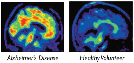
Can
Alzheimer's Disease be seen in the brain during life?
What if you could
help this happen by giving the gift of knowledge for the benefit of others?
UCRT is helping in the
evaluation of a novel imaging tracer, florpiramine F 18 (18F-AV-45) to see if it can
identify the amyloid brain plaques associated with Alzheimer’s Disease.
Until now, these plaques could only be seen by using a microscope to look at
the brain of a patient after they died. Now, using florpiramine,
a new imaging tracer, it may be possible to see amyloid deposits in the brain in living patients
using a PET scan. The purpose of this study is to definitively determine if this
is possible by seeking volunteers who are willing to help achieve this goal by having a florpiramine
PET scan and agreeing to an examination of their brain after they die. The findings
in their ‘brain autopsy’ can then be compared to their PET scan to determine if florpiramine
truly provides a valid measure of the presence or absence of amyloid.
Why is this important?
Alzheimer’s
Disease is the most common cause of dementia, stiking more than 5 million people in the United
States. There is an urgent need to develop a
method to detect its presence before it causes irreversible brain failure. Early
detection will facilitate early treatment. While there has been considerable
progress in understanding how Alzheimer’s Disease develops, only recently has a method been developed that may make
it possible to actually see the pathology in the brains of living patients at the earliest stage of the disease.
By agreeing to participate
in this study of florpiramine, you become part of a team dedicated to turning the dream of a world
free of Alzheimer’s Disease into a reality.
About florpiramine
Florpiramine
is an investigational agent that has not yet been approved by the FDA for use in the routine evaluation of patients. It is not a therapy for dementia or Alzheimer’s Disease. It is a radioactive chemical that when injected into the vein in trace amounts is
carried to the brain where it temporarily binds to amyloid.
The PET scan measures the amount of amyloid in the brain by detecting the amount of florpiramine bound to amyloid. The
goal of this study is to definitively establish that this is true. If this study
demonstrates that a florpiramine PET scan reliably detects amyloid deposits,
it will provide an important new diagnostic tool that physicians can use to more accurately detect Alzheimer’s
Disease in its earliest stage, allowing appropriate treatments to be started at the earliest stage possible and with
greater confidence that the diagnosis is correct.
What happens when a subject joins the study by agreeing to have a florpiramine
PET scan and allow examination of their brain after death?
During the initial research
visit, a member of the study team gathers basic information about the participant, including their medical history and current
medical condition. In addition, they are given a brief (15 to 20 minute) paper
and pencil test to assess their memory and ability to perform simple mental exercises.
They then have a florpiramine PET scan. This procedure takes about 1 hour,
including approximately 15 to 20 minutes lying in the PET scanner while an image of their brain is obtained.
In the weeks following
the PET scan, a member of the study team will maintain periodic contact to monitor the participant’s medical status
and facilitate completion of the brain-only autopsy at the time of death.
Benefits
There is no direct benefit
to participants in this study beyond the knowledge that through their altruism they are helping advance medical knowledge.
How is brain autopsy performed?
The brain is removed by
an opening created in the back of the skull. Afterward, the skull and overlying
skin are returned to their natural position, leaving no visible indications a brain autopsy has been
done.
What happens after the brain is removed?
The brain is placed in
a chemical preservative and sent to the Sun Health Institute in Sun City,
Arizona. There the small sections
needed to compare to the florpiramine PET findings are removed and prepared for analysis. These sections are then sent to Rush University in Chicago where they
are examined under a microscope to look for evidence of amyloid. The
results are compared to the participant’s PET images to determine if florpiramine accurately
marked the presence or absence of amyloid deposits in their brain.
Will brain autopsy interfere with funeral arrangements or having a
open casket viewing?
No. A brain autopsy will not delay or interfere with the plans for an open casket viewing, traditional funeral
services, cremation, or burial. However, the autopsy must be performed within
24 hours from the time of death to provide valid information for this study.
Will there be any cost to the family?
There are no costs to
the family associated with the brain autopsy or with any procedures done in this study.
Do you know someone who has Alzheimer's Disease (AD)
or is terminally ill?
If someone you know has Alzheimer's Disease or is terminally ill, please contact us at Universal
Clinical Research & Technology (UCRT) at 407.574-3801 or 407.574-7361.
For more information on this and other clinical studies, please call Garry or Marla at 407-574-3801.